Neuro Ophthalmology
What Is Neuro-Ophthalmology?
Headache is one of the most common medical complaints throughout human history and affects more than 80% of the population. One of the causes of headaches can be eye-related problems. Patients who complain of headache are often referred to an ophthalmologist by another physician. Neuro-ophthalmology is the medical field that treats headaches caused by eye diseases and focuses on disorders shared by the eye and the nervous system.
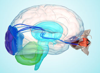
Seeing an object is possible when the image is first formed in the eye and then perceived in the visual center located in the back parts of the brain. If there is a problem along the visual pathways, sudden or progressive vision loss may occur. In disorders affecting eye movements, the main symptom is often double vision, and the problem may involve the eye muscles, the nerves controlling them, or the brain’s control centers.
The following complaints are also within the scope of neuro-ophthalmology:
Temporary vision loss
Double vision
Sudden vision loss
Thyroid-related eye disease
An area in the visual field that cannot be seen (visual field defect)
Eye problems related to multiple sclerosis (MS)
Facial paralysis and facial muscle spasms
Eye problems related to stroke
Migraine and similar eye pain
Drooping eyelid (ptosis)
Differences in pupil size
For accurate diagnosis of head and eye pain, the patient’s history is very important. Success in treatment is possible with a cooperative doctor–patient relationship and regular follow-up.
MS (Multiple Sclerosis) and the Eye
How Does MS Affect the Eye?
Optic neuritis (inflammation of the optic nerve) is an eye condition most commonly seen between ages 18–45, more frequently in women. It typically presents as painful, one-sided vision loss. It occurs in attacks due to an immune system disturbance. In the affected, painful eye, color perception and brightness sensitivity may also decrease.
In about one-third of patients, swelling (edema) occurs at the front part of the optic nerve, while in most cases the involvement is in the part closer to the brain (posterior involvement).
Which Tests Help Diagnose MS?
During diagnosis, the following should be used:
VEP (Visual Evoked Potential)
OCT (Optical Coherence Tomography) to evaluate the nerve fiber layer
Computerized visual field testing
Brain MRI to investigate intracranial lesions
The number and shape of MRI lesions (“spots”) are important in assessing MS risk. MS is a chronic brain disorder that progresses in attacks.
Optic neuritis can be the first initial finding of MS. With treatment, optic nerve inflammation improves substantially within 3–5 weeks, but attacks may recur. The recurrence rate within 10 years is 35%. Optic neuritis is detected at some stage in MS in about 50% of patients, and in 20% it is the first sign of MS.
After optic neuritis, the risk of MS is approximately 30% within the first 5–7 years, and around 75% in women within 15–20 years.
MS attacks may present as optic neuritis, double vision, balance problems, numbness and weakness in the arms or legs.
Current approaches for optic neuritis treatment include high-dose intravenous steroids, and interferon therapy when the likelihood of MS increases.
The course after optic neuritis can be monitored with periodic OCT nerve fiber analyses. If thinning in the OCT nerve fiber layer increases, it may indicate an increase in brain plaques as well. In this condition, follow-up with OCT and VEP is highly important and clinically guiding.
Other Optic Nerve Disorders
Vascular occlusion (intraocular injection may be required)
Toxic optic neuropathy (most commonly due to ethambutol or methanol poisoning; treatable if the patient presents early)
Hereditary optic neuropathy (a new medication has been developed for Leber hereditary optic neuropathy)
Traumatic optic neuropathy (early evaluation is important)
Alzheimer’s and Parkinson’s
What Is the Link Between Alzheimer’s/Parkinson’s and the Eye?
Today, brain diseases such as Alzheimer’s and Parkinson’s, and eye diseases such as glaucoma and macular degeneration (“yellow spot” disease), are considered progressive disorders of nerve tissue (neurodegenerative). Studies have focused on their shared mechanisms, yielding important findings. The OCT test, widely used in diagnosing and treating glaucoma and macular degeneration, is now also applied in Alzheimer’s and Parkinson’s.
Which Tests Support Alzheimer’s and Parkinson’s Diagnosis?
OCT—which provides diagnostic results in comprehensive eye examinations (for glaucoma, macular disease, etc.)—may reveal highly specific findings in Alzheimer’s, Parkinson’s, and mild cognitive impairment. These findings are characterized by damage and thinning in specific regions of the retina or optic nerve fiber layers.
Loss of nerve fibers is important for early diagnosis. OCT may detect nerve fiber thinning before changes become evident in brain regions responsible for memory and movement, enabling earlier diagnosis and treatment opportunities.
In individuals with certain complaints or a family history of Alzheimer’s or Parkinson’s, OCT screening of the retina and optic nerve fibers can play an important role in early detection.
Regular follow-up of diagnosis and treatment using retinal examination methods (FAF and OCT) is important for success. Early diagnosis is possible through retinal evaluation, and treatments aiming to slow disease progression have been initiated in early-detected cases. Due to genetic factors, individuals with a family history of Alzheimer’s or Parkinson’s should have regular eye examinations after age 50.
Optic Nerve Diseases
What Are Optic Nerve Diseases?
Optic Neuritis (Optic nerve inflammation)
Usually affects ages 18–45, more common in women. Typically one-sided, often painful vision loss—especially with eye movements. May occur in attacks due to immune dysregulation. Color and brightness sensitivity decrease. About one-third show swelling at the front of the optic nerve; most involve the part closer to the brain.
In diagnosis, clinics use:
VEP
OCT (nerve fiber imaging)
Contrast sensitivity testing
Computerized visual field examination
Ischemic Optic Neuropathy
Occurs after blockage of small vessels supplying the optic nerve, leading to swelling and bleeding in the nerve. Common in patients with hypertension and diabetes. Intraocular injections may be required.
Toxic Optic Neuropathy
Most commonly seen with ethambutol (used in tuberculosis treatment) and methanol poisoning (e.g., cologne, illicit alcohol). Treatable if the patient presents early.
Hereditary Optic Neuropathy
Leber hereditary optic neuropathy is a maternally inherited bilateral vision loss. A treatment or vision-improving drug has been introduced abroad.
Traumatic Optic Neuropathy
Develops after a direct blow to the eye or head trauma. Early intervention is important.
Tests Performed
Electrophysiology (VEP, ERG, EOG)
FFA (Fluorescein angiography / eye angiography)
OCT (Optic nerve scanning)
VEP (Visual pathways between eye and brain)
Computerized visual field
Contrast sensitivity test
Color vision tests
Prepared by the Dünyagöz Hospital Editorial Board.
Last Updated: 30.05.2023
*This content is for informational purposes only. Please consult your physician for diagnosis and treatment.
Headache is one of the most common medical complaints throughout human history and affects more than 80% of the population. One of the causes of headaches can be eye-related problems. Patients who complain of headache are often referred to an ophthalmologist by another physician. Neuro-ophthalmology is the medical field that treats headaches caused by eye diseases and focuses on disorders shared by the eye and the nervous system.
Seeing an object is possible when the image is first formed in the eye and then perceived in the visual center located in the back parts of the brain. If there is a problem along the visual pathways, sudden or progressive vision loss may occur. In disorders affecting eye movements, the main symptom is often double vision, and the problem may involve the eye muscles, the nerves controlling them, or the brain’s control centers.
The following complaints are also within the scope of neuro-ophthalmology:
Temporary vision loss
Double vision
Sudden vision loss
Thyroid-related eye disease
An area in the visual field that cannot be seen (visual field defect)
Eye problems related to multiple sclerosis (MS)
Facial paralysis and facial muscle spasms
Eye problems related to stroke
Migraine and similar eye pain
Drooping eyelid (ptosis)
Differences in pupil size
For accurate diagnosis of head and eye pain, the patient’s history is very important. Success in treatment is possible with a cooperative doctor–patient relationship and regular follow-up.
MS (Multiple Sclerosis) and the Eye
How Does MS Affect the Eye?
Optic neuritis (inflammation of the optic nerve) is an eye condition most commonly seen between ages 18–45, more frequently in women. It typically presents as painful, one-sided vision loss. It occurs in attacks due to an immune system disturbance. In the affected, painful eye, color perception and brightness sensitivity may also decrease.
In about one-third of patients, swelling (edema) occurs at the front part of the optic nerve, while in most cases the involvement is in the part closer to the brain (posterior involvement).
Which Tests Help Diagnose MS?
During diagnosis, the following should be used:
VEP (Visual Evoked Potential)
OCT (Optical Coherence Tomography) to evaluate the nerve fiber layer
Computerized visual field testing
Brain MRI to investigate intracranial lesions
The number and shape of MRI lesions (“spots”) are important in assessing MS risk. MS is a chronic brain disorder that progresses in attacks.
Optic neuritis can be the first initial finding of MS. With treatment, optic nerve inflammation improves substantially within 3–5 weeks, but attacks may recur. The recurrence rate within 10 years is 35%. Optic neuritis is detected at some stage in MS in about 50% of patients, and in 20% it is the first sign of MS.
After optic neuritis, the risk of MS is approximately 30% within the first 5–7 years, and around 75% in women within 15–20 years.
MS attacks may present as optic neuritis, double vision, balance problems, numbness and weakness in the arms or legs.
Current approaches for optic neuritis treatment include high-dose intravenous steroids, and interferon therapy when the likelihood of MS increases.
The course after optic neuritis can be monitored with periodic OCT nerve fiber analyses. If thinning in the OCT nerve fiber layer increases, it may indicate an increase in brain plaques as well. In this condition, follow-up with OCT and VEP is highly important and clinically guiding.
Other Optic Nerve Disorders
Vascular occlusion (intraocular injection may be required)
Toxic optic neuropathy (most commonly due to ethambutol or methanol poisoning; treatable if the patient presents early)
Hereditary optic neuropathy (a new medication has been developed for Leber hereditary optic neuropathy)
Traumatic optic neuropathy (early evaluation is important)
Alzheimer’s and Parkinson’s
What Is the Link Between Alzheimer’s/Parkinson’s and the Eye?
Today, brain diseases such as Alzheimer’s and Parkinson’s, and eye diseases such as glaucoma and macular degeneration (“yellow spot” disease), are considered progressive disorders of nerve tissue (neurodegenerative). Studies have focused on their shared mechanisms, yielding important findings. The OCT test, widely used in diagnosing and treating glaucoma and macular degeneration, is now also applied in Alzheimer’s and Parkinson’s.
Which Tests Support Alzheimer’s and Parkinson’s Diagnosis?
OCT—which provides diagnostic results in comprehensive eye examinations (for glaucoma, macular disease, etc.)—may reveal highly specific findings in Alzheimer’s, Parkinson’s, and mild cognitive impairment. These findings are characterized by damage and thinning in specific regions of the retina or optic nerve fiber layers.
Loss of nerve fibers is important for early diagnosis. OCT may detect nerve fiber thinning before changes become evident in brain regions responsible for memory and movement, enabling earlier diagnosis and treatment opportunities.
In individuals with certain complaints or a family history of Alzheimer’s or Parkinson’s, OCT screening of the retina and optic nerve fibers can play an important role in early detection.
Regular follow-up of diagnosis and treatment using retinal examination methods (FAF and OCT) is important for success. Early diagnosis is possible through retinal evaluation, and treatments aiming to slow disease progression have been initiated in early-detected cases. Due to genetic factors, individuals with a family history of Alzheimer’s or Parkinson’s should have regular eye examinations after age 50.
Optic Nerve Diseases
What Are Optic Nerve Diseases?
Optic Neuritis (Optic nerve inflammation)
Usually affects ages 18–45, more common in women. Typically one-sided, often painful vision loss—especially with eye movements. May occur in attacks due to immune dysregulation. Color and brightness sensitivity decrease. About one-third show swelling at the front of the optic nerve; most involve the part closer to the brain.
In diagnosis, clinics use:
VEP
OCT (nerve fiber imaging)
Contrast sensitivity testing
Computerized visual field examination
Ischemic Optic Neuropathy
Occurs after blockage of small vessels supplying the optic nerve, leading to swelling and bleeding in the nerve. Common in patients with hypertension and diabetes. Intraocular injections may be required.
Toxic Optic Neuropathy
Most commonly seen with ethambutol (used in tuberculosis treatment) and methanol poisoning (e.g., cologne, illicit alcohol). Treatable if the patient presents early.
Hereditary Optic Neuropathy
Leber hereditary optic neuropathy is a maternally inherited bilateral vision loss. A treatment or vision-improving drug has been introduced abroad.
Traumatic Optic Neuropathy
Develops after a direct blow to the eye or head trauma. Early intervention is important.
Tests Performed
Electrophysiology (VEP, ERG, EOG)
FFA (Fluorescein angiography / eye angiography)
OCT (Optic nerve scanning)
VEP (Visual pathways between eye and brain)
Computerized visual field
Contrast sensitivity test
Color vision tests
Prepared by the Dünyagöz Hospital Editorial Board.
Last Updated: 30.05.2023
*This content is for informational purposes only. Please consult your physician for diagnosis and treatment.